Luciferin, Caged Luciferin And Coelenterazine
Showing all 7 results
Product info
Description
Coelenterazine is a luminophore found in aquatic organisms and is the substrate for many luciferase enzymes, including Renilla reniformis, Gaussia and aequorins. It is commonly used for bioluminescent calcium detection and the monitoring of reporter genes in BRET (Bioluminescence Resonance Energy Transfer), ELISA and HTS methods as well as chemiluminescent detection of superoxide anions and peroxynitrite in cells or tissues.
Product Specifications:
Coelenterazine, native;
2-(p-Hydroxybenzyl)-6-(p-hydroxyphenyl)-8-benzyl-imidazo[1,2-a]pyrazin-3-(7H)-one
MOLECULAR BIOLOGY GRADE
Formua: C26H21N3O3
MW: 423.46 g/mol
Purity: >95% by TLC
Storage/Handling: Store at -20°C, and protect from light. Keep under inert gas.
Soluble in methanol or ethanol (NOT in DMSO).
Note: It is not recommended to store dissolved coelenterazine at -20°C or -70°C. Coelenterazine is a high energy dioxetanone ring structure that will spontaneously decompose even at low temperatures.
PubChem Chemical ID: 2830
Product info
Description
Coelenterazine 400A is a derivative of coelenterazine and is a good substrate for Renilla reniformis luciferase (RLuc), but does not oxidize well with Gaussia (GLuc). It is the preferred substrate for BRET (Bioluminescence Resonance Energy Transfer) since its emission maximum of 400nm has minimal interference with the GFP emission.
Product Specifications:
Coelenterazine 400 a
2,8-Dibenzyl-6-phenyl-imidazo[1,2a]pyrazin-3-(7H)-one
DeepBlueCTM
Formula: C26H21N3O
MW: 391.48 g/mol
Purity: >95% by TLC
Color: Yellow to orange powder
Storage/Handling: Store at -20°C, and protect from light. Keep under inert gas.
Soluble in methanol or ethanol (NOT in DMSO).
Note: It is not recommended to store dissolved coelenterazine 400a at -20°C or -70°C. Coelenterazine 400a is a high energy dioxetanone ring structure that will spontaneously decompose even at low temperatures.
PubChem CID: 135750016
Product info
Description
Tested and Certified for in vivo imaging (See “Luciferin FAQ” in additional information)
Luciferin is a common bioluminescent reporter used for in vivo imaging of the expression of luciferase. This substrate for the firefly luciferase enzyme utilizes ATP and Mg2+ as cofactors to emit a characteristic yellow-green emission in the presence of oxygen, which shifts to red light in vivo at 37°C. Through the utilization of ATP, the reaction can be further used to indicate the presence of energy or life in order to function as a life-death stain.
Luciferin is a common reagent used throughout the biotechnology field and specifically for in vivo imaging. Luciferase labeled tumor cells, stem cells or infectious diseases are often inoculated into research animals such as rats or mice for investigation. The injection of luciferin allows for the real-time, noninvasive monitoring of disease progression and/or drug efficacy in these model systems through Bioluminescence Imaging (BLI).
Luciferin is also commonly used for in vitro research, including luciferase and ATP assays, gene reporter assays, high throughput sequencing and various contamination assays.
Firefly Luciferin is identical to Beetle Luciferin. (See “Additional Information” for structures.)
Product Specifications:
D-Luciferin Firefly, free acid
4,5-Dihydro-2-(6-hydroxy-2-benzothiazolyl)-4-thiazolecarboxylic acid
MOLECULAR BIOLOGY GRADE
Formula: C11H8N2O3S2
MW: 280.33 g/mol
Color: Off white to light yellow powder
Purity: >99% pure
Storage/Handling: Store desiccated at -20°C. Protect from light.
PubChem Chemical ID: 92934
Note: The Free Acid form of luciferin will not dissolve in water unless a dilute base such as NaOH, KOH, sodium bicarbonate or potassium bicarbonate is added to adjust the pH. The potassium or sodium salts of luciferin are more convenient for experiments, particularly for in vivo imaging, since they readily dissolve in water or buffer.
Product info
Description
Tested and Certified for in vivo imaging (See “Luciferin FAQ” in additional information)
Luciferin is a common bioluminescent reporter used for in vivo imaging of the expression of luciferase. This water soluble substrate for the firefly luciferase enzyme utilizes ATP and Mg2+ as cofactors to emit a characteristic yellow-green emission in the presence of oxygen, which shifts to red light in vivo at 37°C. Through the utilization of ATP, the reaction can be further used to indicate the presence of energy or life in order to function as a life-death stain.
Luciferin is a common reagent used throughout the biotechnology field and specifically for in vivo imaging. Luciferase labeled tumor cells, stem cells or infectious diseases are often inoculated into research animals such as rats or mice for investigation. The injection of luciferin allows for the real-time, noninvasive monitoring of disease progression and/or drug efficacy in these model systems through Bioluminescence Imaging (BLI).
Luciferin is also commonly used for in vitro research, including luciferase and ATP assays, gene reporter assays, high throughput sequencing and various contamination assays.
Firefly Luciferin is identical to Beetle Luciferin. (See “Additional Information” for structures.)
Product Specifications:
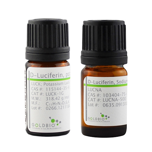
D-Luciferin, Potassium Salt,
4,5-Dihydro-2-(6-hydroxy-2-benzothiazolyl)-4-thiazolecarboxylic acid potassium salt,
MOLECULAR BIOLOGY GRADE (Premium Pure)
Formula: C11H7N2O3S2K
MW: 318.42 g/mol
Color: Light yellow powder
Purity: >99% pure. (Quality verified by nine independent criteria including HPLC and FTIR.)
Storage/Handling: Store desiccated at -20°C. Protect from light.
PubChem Chemical ID:
44134804
For the full recipe and instructions, go to GoldBio’s Luciferin in vitro Handbook.
Product info
Description
Tested and Certified for in vivo imaging (See “Luciferin FAQ” in additional information)
Luciferin is a common bioluminescent reporter used for in vivo imaging of the expression of luciferase. This water soluble substrate for the firefly luciferase enzyme utilizes ATP and Mg2+ as cofactors to emit a characteristic yellow-green emission in the presence of oxygen, which shifts to red light in vivo at 37°C. Through the utilization of ATP, the reaction can be further used to indicate the presence of energy or life in order to function as a life-death stain.
Luciferin is a common reagent used throughout the biotechnology field and specifically for in vivo imaging. Luciferase labeled tumor cells, stem cells or infectious diseases are often inoculated into research animals such as rats or mice for investigation. The injection of luciferin allows for the real-time, noninvasive monitoring of disease progression and/or drug efficacy in these model systems through Bioluminescence Imaging (BLI).
Luciferin is also commonly used for in vitro research, including luciferase and ATP assays, gene reporter assays, high throughput sequencing and various contamination assays.
Firefly Luciferin is identical to Beetle Luciferin. (See “Additional Information” for structures.)
Product Specifications:
D-Luciferin, Sodium Salt
4,5-Dihydro-2-(6-hydroxy-2-benzothiazolyl)-4-thiazolecarboxylic acid sodium salt
MOLECULAR BIOLOGY GRADE (Premium Pure)
Formula: C 11H9N2O4S2Na
MW: 320.32 g/mol
Color: Light yellow powder
Purity: >99% pure (Quality verified by nine independent criteria including HPLC and FTIR.)
Storage/Handling: Store desiccated at -20°C. Protect from light.
PubChem Chemical ID: 23710675
For the full recipe and instructions, go to GoldBio’s Luciferin in vitro Handbook.
Product info
Description
Tested and Certified for in vivo imaging (See “Luciferin FAQ” in additional information)
Luciferin is a common bioluminescent reporter used for in vivo imaging of the expression of the luciferase . This substrate for the firefly luciferase enzyme utilizes ATP and Mg2+ as cofactors and emits a characteristic yellow-green emission in the presence of oxygen, which shifts to red light in vivo at 37°C.
Luciferin is a common reagent used throughout the biotechnology field and specifically for in vivo imaging. Luciferase labeled tumor cells, stem cells or infectious diseases are often inoculated into research animals such as rats or mice for investigation. The injection of luciferin allows for the real-time, noninvasive monitoring of disease progression and/or drug efficacy in these model systems through Bioluminescence Imaging (BLI).
DMNPE-caged luciferin can be used to measure intracellular functions since it readily crosses cell membranes. Once it is inside the cell, active luciferin can be released by UV light or by the action of endogenous intracellular esterases. DMNPE-caged luciferin makes it easier to follow the changes in gene expression in live cells. It can also be used in combination with DMNPE-caged ATP.
Product Specifications:
DMNPE-caged Luciferin
D-Luciferin 1- (4,5-dimethoxy-2- nitrophenyl) ethyl ester
MOLECULAR BIOLOGY GRADE
Color: Light yellow powder
Formula: C21H19N3O7S2
MW: 489.53 g/mol
Soluble in DMSO or DMF.
Storage: Store desiccated at -20°C, protect from light.
PubChem CID: 2762658
Product info
Description
Tested and Certified for in vivo imaging (See “Luciferin FAQ” in additional information)
Luciferin is a common bioluminescent reporter used for in vivo imaging of the expression of luciferase. This water soluble substrate for the firefly luciferase enzyme utilizes ATP and Mg2+ as cofactors to emit a characteristic yellow-green emission in the presence of oxygen, which shifts to red light in vivo at 37°C. Through the utilization of ATP, the reaction can be further used to indicate the presence of energy or life in order to function as a life-death stain.
Luciferin is a common reagent used throughout the biotechnology field and specifically for in vivo imaging. Luciferase labeled tumor cells, stem cells or infectious diseases are often inoculated into research animals such as rats or mice for investigation. The injection of Luciferin allows for the real-time, noninvasive monitoring of disease progression and/or drug efficacy in these model systems through Bioluminescence Imaging (BLI).
Luciferin is also commonly used for in vitro research, including luciferase and ATP assays, gene reporter assays, high throughput sequencing and various contamination assays.
L-Luciferin is the chiralic sister to D-luciferin. While most researchers claim that L-luciferin does not produce any light, Lembert has reported that it produces a very weak and extremely slow manifesting light (Lembert, 1996). Nakamura suggested that an enzyme system incorporating luciferase might be responsible for changing the chirality of the L-luciferin to D-luciferin (Nakamura et al., 2006) (See Reference Library).
Product Specifications:
L-Luciferin, Potassium Salt
4,5-Dihydro-2-(6-hydroxy-2-benzothiazolyl)-4-thiazolecarboxylic acid potassium salt
Forms a yellow, clear solution when dissolved 1% in water.
Formula: C11H7N2O3S2K
MW: 318.42 g/mol
Color: Light yellow powder
Purity: >98% by HPLC
Storage/Handling: Store desiccated in -20°C, and protect from light.
PubChem CID: 135845006